
Are we really still impressed by high-resolution images? In an age of simulated realities and AI-generated everything, the idea of painstakingly creating detailed 3D models of…ants…feels almost quaint. But the Antscan project, which has produced stunningly precise images of over 700 ant species, isn’t about spectacle. The real story here isn’t the pretty pictures—it’s the quiet revolution happening in how we do science, and the surprising places that revolution is taking root.
Beyond the Bug: The Power of Synchrotron Light
For years, biologists have relied on dissection and limited microscopy to understand the inner workings of insects. It’s a destructive process, offering only snapshots of a static organism. Julian Katzke, a postdoc at the Smithsonian National Museum of Natural History and key researcher on Antscan, and his team took a different approach. They didn’t slice and dice; they saw through. Using a synchrotron x-ray source in Germany – essentially a giant particle accelerator repurposed for imaging – they scanned preserved ant specimens, building up a 3D model with a resolution of 1.22 micrometers. To put that in perspective, that’s small enough to distinguish individual muscle fibers and the tiny hairs covering an ant’s body. This wasn’t a simple task. It required a robotic vial-swapping system to feed hundreds of specimens into the x-ray beam, a logistical feat as impressive as the imaging itself.
Original reporting: sciencefriday.com.
The synchrotron isn’t some niche academic tool. It’s a massive investment, typically used for materials science and physics. The fact that it’s now being applied to entomology speaks volumes about the growing accessibility – and the increasing sophistication – of biological imaging. It’s a shift driven by the realization that understanding complex systems requires seeing them in their entirety, not just in fragments. And it’s a shift that’s going to ripple far beyond the study of ants.
From Museum Collections to Digital Archives
What’s particularly compelling about Antscan is its reliance on existing museum collections. Researchers didn’t need to embark on massive, potentially disruptive field expeditions. They tapped into a wealth of already-collected specimens, effectively breathing new life into decades-old research. This is a crucial point. The cost of new scientific discovery is skyrocketing, and the logistical hurdles of fieldwork are becoming increasingly difficult to overcome. Digitizing existing resources – and finding innovative ways to analyze them – is becoming a cornerstone of modern biology.
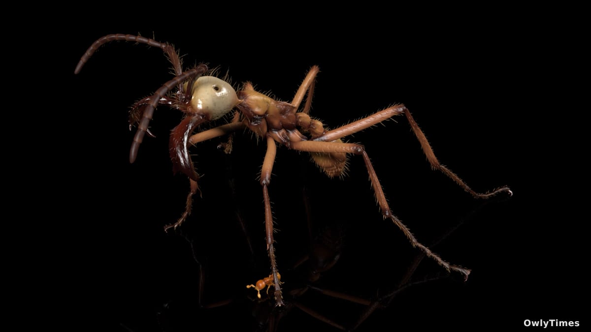
The resulting data isn’t just for scientists, either. The Antscan team is actively exploring artistic applications, creating visually arresting 3D renderings of ant anatomy. These aren’t sterile scientific visualizations; they’re evocative representations of incredibly complex organisms, designed to inspire wonder and appreciation. The images, showcasing everything from the digestive tracts to the intricate brain structures, are available for public viewing, blurring the lines between research and outreach.
Why This Matters to You (Even If You Hate Bugs)
You might be wondering why anyone should care about high-resolution images of ants. After all, most people’s interactions with ants involve annoyance or a quick squish. But ants are ecological powerhouses, playing critical roles in soil health, pollination, and seed dispersal. Understanding their anatomy and physiology is crucial for understanding entire ecosystems. More broadly, the techniques developed for Antscan have implications for medical imaging, materials science, and even robotics.
Consider the potential for non-destructive analysis of delicate artifacts, or the development of bio-inspired robots that mimic the incredible strength and agility of ants. The precision achieved by Antscan could also inform the design of new micro-devices and sensors. This isn’t about glorifying insects; it’s about leveraging the power of advanced technology to unlock secrets hidden within the natural world – secrets that could ultimately benefit us all.
The Future of Seeing: Predictive Modeling and AI
The Antscan project is a proof of concept, demonstrating the power of combining advanced imaging techniques with robotic automation. But it’s just the beginning. The real leap forward will come when we start using artificial intelligence to analyze this vast trove of 3D data. Imagine AI algorithms capable of identifying subtle anatomical differences between ant species, predicting their behavior, or even designing new materials based on their unique structures.
Within the next five years, I predict we’ll see the emergence of “digital twins” of entire ecosystems, built from data like that generated by Antscan. These virtual worlds won’t just be visually stunning; they’ll be powerful tools for predicting the impact of climate change, invasive species, and other environmental threats. The question isn’t whether we can create these digital ecosystems, but whether we’ll have the foresight to use them responsibly.